Laser microdissection: Better research for cancer

Early detection of cancer can save lives. Laser microdissection enables specific cell examinations and precise cancer research. Experts from MicroDissect GmbH are developing innovative membrane-supported consumables for this purpose – a guarantee for samples with high purity and reliable analysis results, and thus an important basis for molecular biology.

Eine sich teilende Krebszelle
Malignant tumors are called carcinomas. The cancer cells of a carcinoma divide in an uncontrolled manner - which means healthy neighboring tissue can be attacked and destroyed. (Credit: Giovanni Cancemi / shutterstock.com)

The earlier a tumor is detected, the more targeted the treatment can start – which also increases the chances of survival. In order to investigate the causes of cancer, scientists examine the abnormal cells, for example, in a biopsy. The physician takes a tissue sample from the patient and examines it for cell changes, such as those that occur in cancer. The exact classification of the cells then determines the further prognosis. Only a tissue analysis can tell with certainty whether the mutation is benign or malignant.

Tissue samples in precision

Tumors usually consist of a heterogeneous mixture of cells. However, it is often not the size of the tumor that is relevant, but rather, it is the tumor’s aggressiveness. This characteristic can be clarified even more precisely using laser microdissection. The method enables the analysis of individual cells using light microscopy. For this purpose, a wafer-thin tissue section is viewed under the microscope. The laser enables the analysis of subcellular genes, RNA and proteins. “So far, there have been no alternatives. Researchers have tried to carry out the isolation manually,” explains Dr. Dorina Böhm, Managing Director of MicroDissect GmbH, a manufacturer of consumables for membrane-supported microdissection. With the developed laser system, the desired cell segment can be marked on the microscope slide by computer and separated from undesired cell segments without contact. Electronically controlled separation thus offers scientists the opportunity to obtain microscopically small and highly pure samples. “Since the 1990s, laser microdissection has established itself as a standardized method and has been continuously further developed ,” says Böhm.

“A chance for personalized medicine”

Not all cancers are the same. In cases of this disease, classification of the underlying mutation plays an important role. For more than 18 years, laser microdissection (LMD) has helped doctors to find different causes, and thus effective treatment methods, for cancer patients. Dr. Falk Schlaudraff and Dr. Christoph Greb from Leica Microsystems explain why their LMD microscopes contribute to cancer research and even more scientific fields.

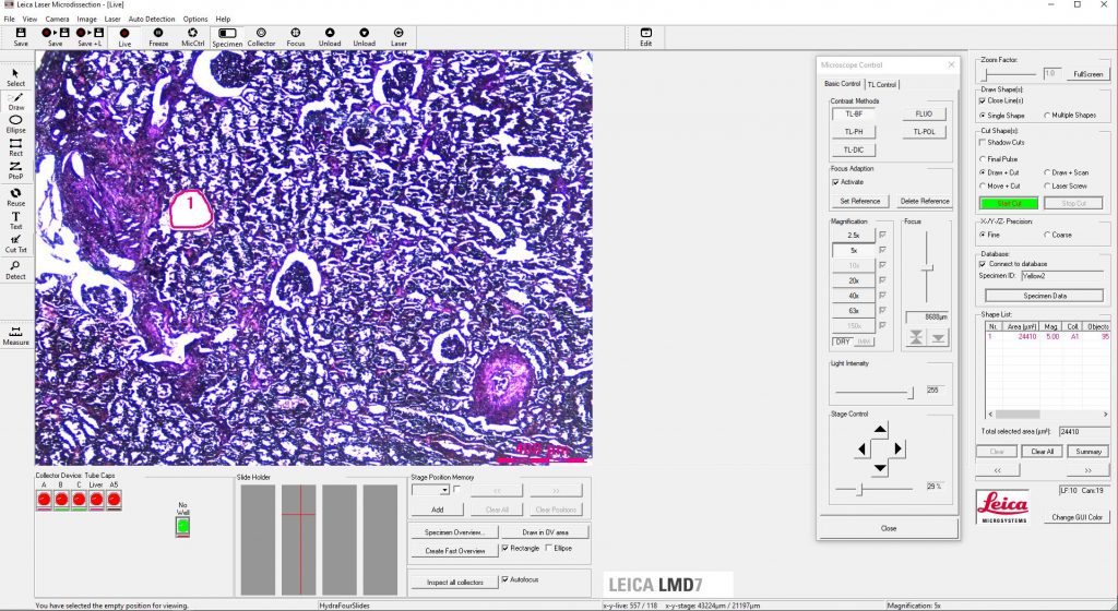
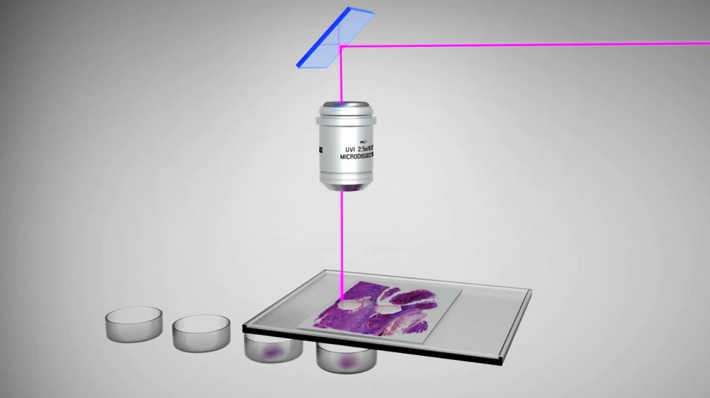
Schlaudraff: The classical field of application is usually pathology, especially tumor research. Laser microdissection enables the controlled, precise isolation of diseased cells from a heterogeneous tissue sample in order to obtain a pure dissection for follow-up examinations. This allows more precise conclusions to be drawn: The results of the investigations are not based on a mixture of healthy and diseased cells, but they can be specifically assigned to one or the other population.

Greb: Laser microdissection also plays an important role in the neurosciences and is used to separate certain brain areas down to individual neurons. Forensics is a more exotic field of application. LMD enables the separation of biological material of the perpetrator from that of the victim. The resulting genetic fingerprint then often contributes to the capture and identification of the perpetrator. Biologists use laser-assisted microdissection to examine the annual rings of trees. This allows conclusions to be drawn about how the climate must have been about 1,000 years ago. Polar researchers use the method to isolate bacteria from drilling samples of deep ice layers. The creative and varied use of laser microdissection remains unlimited.

Schlaudraff: Leica Microsystems mainly serves the scientific market. Our customers are primarily active in research. But hospitals also use this method for diagnosis. We are active worldwide and sell our instruments in nearly all markets. We are constantly developing these further.

Greb: By the way – in addition to cutting out “dead” areas from tissue sections, scientists can also use our LMD to separate living cells from cell cultures. This application is becoming increasingly popular.

Schlaudraff: In my opinion, the medicine of the future is personalized medicine. The recognition and knowledge of the different types of cancer have strongly influenced treatment methods. LMD offers the opportunity to identify causes and develop coordinated forms of therapy for the patient.

Greb: At the same time, the analysis techniques based on the results of laser microdissection have also become increasingly precise. Previously, it was necessary to concentrate on an entire tissue sample; with the aid of LMD, individual cells can now be separated if required. This enables users to exploit the full potential of molecular biological analysis methods. I therefore greatly appreciate the uniqueness of our upright laser microdissection systems. The use of gravity in our LMD method offers many advantages for the pure and contactless isolation of numerous dissections.

Optimizing qualitative standards

But success does not come without challenges: Conventional microscope slides are unsuitable for the further examination of tissue. A single cell can hardly be isolated from a glass slide without being damaged. And any manual intervention with medical cutlery endangers the goal of high-purity sample collection. “Only absolutely pure cell samples allow conclusions to be drawn about the molecular genetic causes of a malfunction and the development of a tumor,” Böhm explains. Thus, the inert membrane from MicroDissect offers a simple and efficient solution for further sample analyses. The membrane-supported laser microdissection is based on the inert nature of the carrier membrane.

In addition to a modern laser system, optimally adapted medical accessories are a prerequisite for the high purity of tissue samples. The consumables needed for this purpose are produced by MicroDissect GmbH, based in Herborn, Central Hessen. The company produces them under sterile room conditions of the highest quality. “When the dissectate has been isolated by the laser beam, it falls into the lid of a so-called cap. The optical element contained in the cap makes the cell sample visible under the microscope,” says Böhm. The highly pure membrane, which is free of grease and adhesives, acts like a tablet, but does not interfere with further examination.

"Since the 1990s, laser microdissection has established itself as a standardized method and has been continuously further developed."
Dr. Dorina Böhm
Dr. Dorina Böhm
Geschäftsführerin der MicroDissect GmbH

An invention in science

Still, it is hard to imagine cancer research without membrane-supported laser microdissection. Stem cell researchers are also currently using the laser system to expand their dissection possibilities. They expect progress in regenerative medicine and stem cell therapy. And even plant researchers and botanists are hoping that modern isolation technology will lead to new insights into parasites and pathogens, the results of which could be decisive for the agriculture of the future. For these research possibilities, the experts at MicroDissect respond flexibly to customer requirements. This includes the production of development samples as well as customer-specific small series and OEM products. But membrane-supported laser microdissection is already contributing to the precise research of tumor cells – and helping to improve the lives of cancer patients.

company profile

MicroDissect GmbH

MicroDissect GmbH, located in the Lahn-Dill region since 2001, manufactures and supplies consumables for membrane-supported microdissection. Its customers include well-known suppliers of systems for laser microdissection and well-known universities, institutes and laboratories.

Leave a Comment